Diagnostic testing is an important step in working towards finding the right treatment for a patient. Through testing, doctors and other medical professionals can accurately diagnose any disease and determine the appropriate type of treatment to cure or manage it. Advances in medical technology have allowed tests to become more sophisticated, making earlier detection and treatment possible.
Today, technology has allowed the fusion of imaging procedures such as PET scan and CT scan to be performed together.
What is a PET-CT Scan?
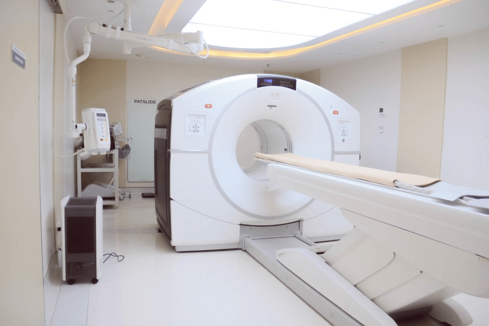
Positron Emission Tomography (PET) and Computerized Tomography (CT) are standard radiological imaging tools which doctors use to view the body and its functions.
A PET scan will involve injecting a special radioactive tracer that will settle in certain areas of the body, making it easier to view specific conditions Meanwhile, a CT scan uses special X-ray equipment to produce 3D images of the body and its inner structures.
A PET-CT scan produces fused images, allowing doctors to correlate and interpret data from two different imaging techniques in one image. This means more precise information, leading to a more accurate diagnosis overall.
Due to their precision and accuracy, PET-CT scans are often performed for the following reasons:
- Diagnosis and detection of cancer
- Determining the spread of cancer cells in the body
- Assessment of tissue metabolism and viability
- Evaluation of neurological abnormalities like tumors, seizures, and memory disorders
- Observation of the effects of myocardial infarction on areas of the heart
- Identification of heart muscle areas needing angioplasty or coronary artery bypass surgery
- Planning the appropriate treatment for a disease
The PET-CT Scan Process
Prior to the exam, a limited carbohydrate diet will be put into place 24 hours before the scheduled appointment.
Patients are discouraged from eating or drinking anything except water six hours before the exam. This is to prevent possible alterations in the distribution of the tracer in the body, leading to an inaccurate scan. For individuals with diabetes, doctors may give special instructions to prepare for the scan.
On the day of the procedure, patients will be asked to remove any jewelry and other metal objects to avoid any interference with the scanner. A radioactive tracer will be injected through a cannula. This will take around an hour to spread throughout the body and settle into the tissues.
Patients will then be asked to lie flat on their back on the machine bed which will slowly slide backward and forward through the scanner to capture the images. The patient must remain still while the scan is ongoing to capture clear images. While the scan itself is painless, discomfort may arise from the lack of movement. The full scan will take approximately 20 to 30 minutes.
Depending on the area for examination, additional views may be required. This can extend the procedure time to 3 hours.
After the scan, the Nuclear Medicine physician and radiologist will review the images in case more scans are needed to clarify or better visualize certain areas. Unless instructed by a medical professional, the patient may resume normal activities after the scan.
Increased fluid intake is encouraged to help flush out the tracers. It is also recommended for patients to limit interaction with pregnant women and infants to avoid exposing them to radiation. Bruising or swelling may occur on the injection site.
Risk Factors
Like any other medical procedure, a PET-CT scan has its share of risks. Further consultation with doctors is needed to better assess the risks for individuals with the following conditions:
The radioactive tracer contains only minute amounts of radiation, which is generally harmless to most people. However, this radiation can pose a risk to the development of unborn babies. It is crucial to consult with a doctor for pregnancy-safe alternatives to a PET-CT scan.
Exposure to the radioactive tracer can affect the milk of breastfeeding mothers. Expressing milk for at least one feed prior to the exam is encouraged, as breastfeeding will need to be delayed until the tracer is completely flushed out.
Sometimes, patients are given both radioactive tracer (PET portion) and contrast (CT portion). For some patients undergoing PET-CT studies, there is a risk of allergy to iodinated contrast. Persons with pre-existing conditions and individuals taking beta-blockers and nonsteroidal anti-inflammatory drugs (NSAIDs) may need additional consultation before undergoing a scan.
Scans Save Lives
Diagnostic testing is necessary for detecting and treating illnesses. A PET-CT scan provides a combination of the two separate exams, allowing doctors to get a clearer and more accurate look at the body’s overall condition. This leads to a better, more accurate diagnosis and higher chances of recovery.
For quality diagnostic testing and treatment, Makati Medical Center offers top-of-the-line PET-CT scanning & radiology services.

